Most and Least Nutritive Fruits In World Source: Pixabay Most and Least Nutritive Fruits An analysis of the 38 commonly eaten raw (as opposed to dried) fruits shows that the one with the highest calorific value is the avocado (Persea americana) with 741 calories per edible lb. That with the lowest value is cucumber with 73 calories per lb. Avocados probably originated in Central and South America and also contain vitamins A, C. and E and 2.2% protein. Biggest Apple An apple weighing 3 lb 1 oz was reported by V. Loveridge of Ross-on Wye, England in 1965. Largest Artichoke An 8-lb artichoke was grown in 1964 at Tollerton, N Yorkshire England, by A. R. Lawson Largest Broccoli A head of broccoli weighing 28 lb 14 3/4 oz was grown in 1964 by J. T. Cooke of Huntington, W. Sussex, England. Largest Cabbage In 1865 William Collingwood of The Stalwell, County Durham, England, grew a red cabbage with a circumference of 259 in. It reputedly weighed 123 lb. Largest Carrot A carrot weighing 11 ...
Electricity and bioelectricity :DIGITAL SUBTRACTION ANGIOGRAPHY, COMPUTED TOMOGRAPHY,MAGNETIC RESONANCE IMAGING
Electricity and bioelectricity :DIGITAL SUBTRACTION ANGIOGRAPHY, COMPUTED TOMOGRAPHY, MAGNETIC RESONANCE IMAGING
DIGITAL SUBTRACTION ANGIOGRAPHY
The femoral artery is a commonly used entry point and is accessed at the level of the femoral head. At this location, its position is relatively superficial and hemostasis can therefore be achieved with ease because the artery can be compressed against the femoral head once the procedure is completed Different catheters are used to access the branch arteries requiring imaging Once the catheter is in the appropriate vessel iodinated contrast (often called "X-ray dye'') is injected at a controlled rate and volume either via a power injector or by hand injection.
Imaging can be acquired on film radiographs (which are processed similarly to routine radiographs) or much more commonly with digital subtraction angiography (DSA). In DSA, images are processed with the help of a computer The initial image has no contrast and is called the "mask." The X-ray images are then obtained in rapid sequence while contrast is injected. The computer then digitally "subtracts" the mask from the subsequent images leaving only the contrast, thereby yielding finely detailed imaging of the vasculature.
If an area of stenosis or narrowing is identified, it can be treated using percutaneous transluminal angioplasty (PTA), with or without the use of a metallic stent.
If blood flow cessation is required in an area of bleeding (eg, after trauma) or as preoperative embolization to reduce operative blood loss at subsequent surgery, a catheter can be advanced to the vessel requiring embolization Various materials can be delivered into the artery to stop flow Embolization materials include metallic coils, polyvinyl alcohol particles (PVA, which is fixed-size particulate material), alcohol, chemotherapeutic material, autologous clot, biocompatible glue, and other agents.
COMPUTED TOMOGRAPHY
CT Imaging Orientation
 |
| Source: Pixabay |
Computed tomography (CT) uses ionizing radiation to create an image. This allows visualization of a greater variety of tissue structures beyond the four basic densities (air, bone, soft tissue, and fat) that are seen on a conventional radiograph Unlike conventional x-rays, which utilize one projection to form an image. CT uses multiple small projections across the body and combines the information to form the image It is this combining of the images that allows greater soft tissue detail to be displayed Each individual picture of a computed tomography (CT) study is referred to as a section or an axial "slice." This is because the picture must be interpreted as if the patient has been completely sectioned in an axial plane, like a loaf of bread, with the
viewer looking at the section from the feet towards the head The amount of the x-ray beam that a particular voxel of tissue attenuates can be represented by a number called the Hounsfield.
Contrast Studies
Intravenous contrast as well as oral and sometimes rectal contrast agents may be used in CT scans. The small "+C" label in the alphanumeric image of each section indicates that intravenous contrast was used. Also, if the aorta, kidneys, or ureter is radiopaque or white, it is a good indication that I contrast was used If the stomach or small or large bowel is radiopaque, oral contrast has been given.
MAGNETIC RESONANCE IMAGING
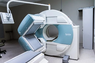
Magnetic resonance imaging (MRI ) has its greatest application in the fields of neuroradiology and musculoskeletal radiology
 |
| Source: Pixabay |
To form a magnetic resonance image the patient is placed in a strong uniform magnetic field. The magnetic field aligns hydrogen nuclei within the patient in the direction of the field. The nuclei are disturbed from this orientation by application of an external radiofrequency (RF) pulse After the RF pulse is stopped the hydrogen nuclei return to their alignment within the externally imposed magnetic field, giving off RF signals as they lose energy.
The frequency of the RF signal emitted from the hydrogen nucleus as it returns to its orientation within the field is determined by the strength of that field. Therefore, the location of the RF the RF signal given off by each hydrogen nucleus can be calculated Each RF signal is analyzed by the computer for its intensity and other criteria.
The signals are then assigned grayscale values (white to black) on the detector by the computer Since this process of creating an image based on tissue characteristics is completely different from the absorption of x-rays by different tissues, MR images can show different types of pathologies and hence its utility For example, MRI can discriminate soft tissue differences better than CT scans and is often used to define soft tissue abnormalities like herniated discs, ligament tears, and soft tissue tumors in the spine.
There are two basic sequences in MRI that are important to understand and recognize
1 Tl-weighted
2. T2-weighted sequences
The "weighting" represents the exploitation of specific properties of hydrogen atoms that are exposed to a magnetic field T1-weighted images classically demonstrate water as hypointense (dark) and fat as hyperintense (bright), with different soft tissues expressed as a gradient in between In T2-weighted images, water is represented as hyperintense and fat as hypo intense (again with soft tissues in the middle). Many of the more complicated sequences (gradient echo, FLAIR, etc.) are based on these basic sequences.
Advantages and Disadvantages of MRI
MRI's clear advantage is that it uses no ionizing radiation. Disadvantages of MRI are that it is generally more costly than CT, is less available, and takes longer to perform Another disadvantage is the inability to scan patients who have ferromagnetic material such as some shrapnel in them.
These metallic fragments can actually move with the magnetic field and cause the patient significant discomfort or damage Although MRI-compatible pacemakers are in development, generally speaking pacemakers are contraindicated because of their ferromagnetic properties, the MRI can heat the leads and inappropriately trigger the pacemaker. But some metal, based on location, type, or duration in place, may not contraindication to an MRI.

Comments
Post a Comment